Biliary PoCUS: What to Look for in Right Upper Quadrant Pain
Biliary point-of-care ultrasound can provide valuable bedside information in patients with right upper quadrant pain. A focused scan helps clinicians identify gallstones and look for findings that support acute cholecystitis.
Right upper quadrant pain? Grab the probe.
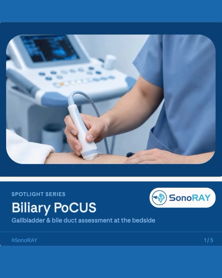
Ultrasound is generally the preferred initial imaging test when biliary disease is suspected. At the bedside, biliary PoCUS can rapidly narrow the differential and guide the next steps in evaluation. Begin by scanning the entire gallbladder in both long and short axes.
Gallstones usually appear as bright, echogenic structures within the gallbladder. They commonly settle in dependent areas, move when the patient is repositioned, and produce clean posterior acoustic shadowing.
Then look for signs of inflammation:
- A probe-directed sonographic Murphy sign
- Gallbladder wall thickening
- Pericholecystic fluid
- Gallbladder distension
- Common bile duct dilation when obstruction is suspected
These findings become more meaningful when they occur together and match the patient’s history, examination, and laboratory results.
Gallstones alone indicate cholelithiasis and do not automatically confirm acute cholecystitis. Some patients have gallstones without symptoms, while acute cholecystitis is suggested by a combination of stones, inflammatory ultrasound findings, and an appropriate clinical presentation.
PoCUS can accelerate clinical decision-making, but a negative or technically limited bedside examination does not completely exclude biliary disease. Evidence suggests clinician-performed PoCUS has high specificity but more moderate sensitivity for acute cholecystitis, so further imaging may still be required when clinical concern remains high.
Find the gallbladder.
- Follow it from fundus to neck.
- Interpret the findings in clinical context.
About the Author
Written by SonoRAY (@admin)