
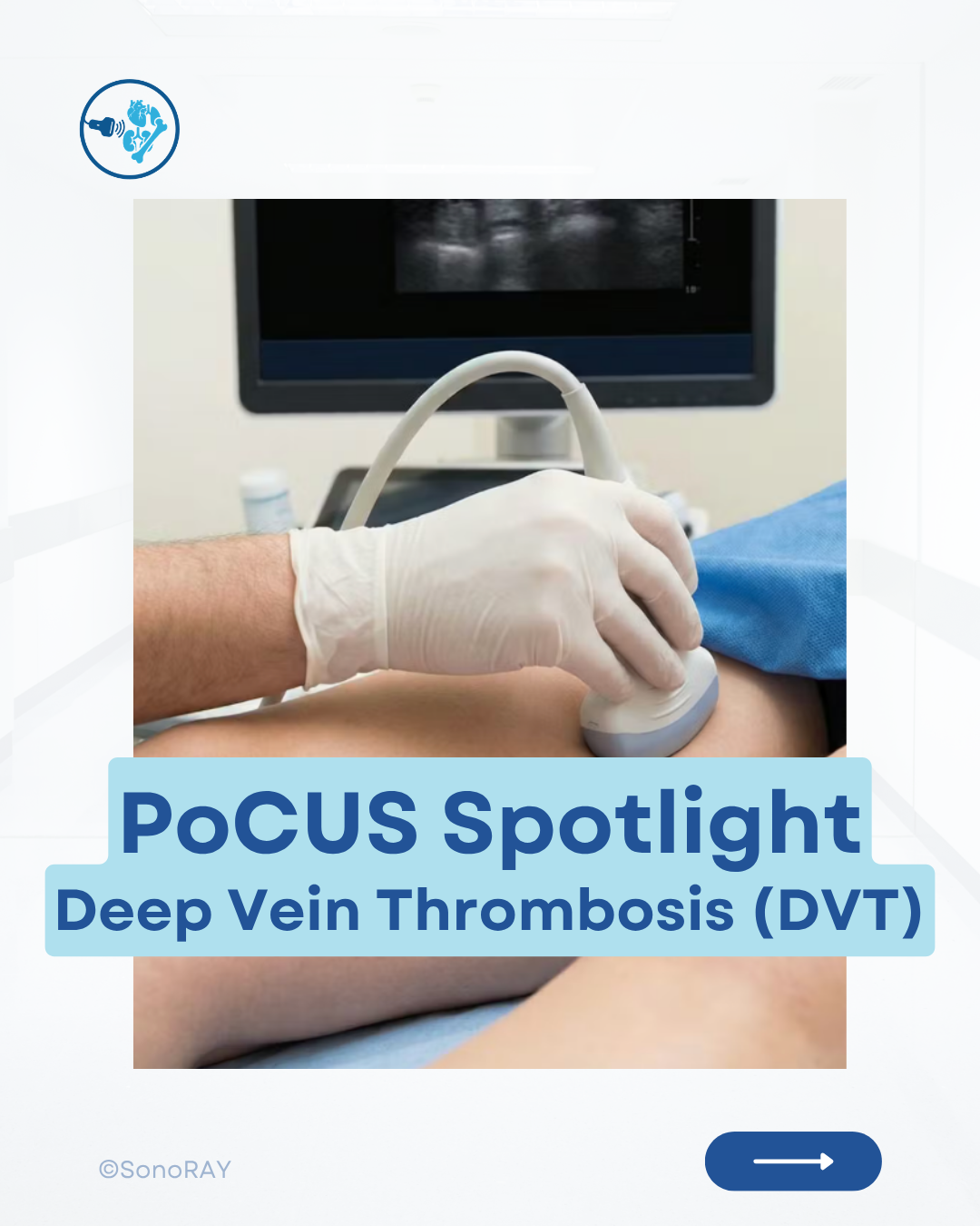
DVT PoCUS: Recognizing a Non-Compressible Vein
Deep vein thrombosis is a time-sensitive diagnosis that can lead to pulmonary embolism. Compression PoCUS allows clinicians to rapidly assess the proximal lower-extremity veins for evidence of thrombosis at the bedside.
Unilateral leg swelling or pain? Consider DVT.
Deep vein thrombosis occurs when a blood clot forms within a deep vein, most commonly in the lower extremity. Early recognition matters because part of the clot may migrate to the lungs and cause a potentially life-threatening pulmonary embolism.
Compression point-of-care ultrasound can rapidly evaluate the proximal lower-extremity veins at the bedside, as demonstrated in Sonoray’s point-of-care ultrasound education.
Begin with the patient supine and the affected leg slightly externally rotated. Use a high-frequency linear transducer and examine the veins in the transverse plane.
Key regions commonly assessed include:
- The common femoral vein
- The femoral vein
- The popliteal vein
Follow the venous system distally and apply firm, perpendicular pressure at short intervals. A normal vein should collapse completely, allowing its anterior and posterior walls to touch.
The Key Finding: Non-Compressibility
The most important ultrasound finding of DVT is a vein that does not fully compress. Thrombus may also appear as echogenic material within the vessel, but a fresh clot can be relatively anechoic and difficult to see. For this reason, the diagnosis should be based primarily on compressibility rather than clot visualization alone.
Additional findings may include:
- Venous distension
- Abnormal or absent colour flow
- Reduced Doppler phasicity
- Visible intraluminal material
These findings can support the diagnosis, but they do not replace systematic compression.
A negative limited PoCUS examination does not exclude every DVT. Isolated calf, pelvic or intervening femoral-vein thromboses may be missed by a restricted scanning protocol, highlighting the importance of appropriate training such as an Acute Care POCUS Course. When clinical concern remains high, formal imaging, D-dimer testing or repeat ultrasound may still be required according to the patient’s pretest probability and local pathway.
Patients with suspected DVT who also develop chest pain, shortness of breath, syncope or unexplained hypoxia require immediate assessment for pulmonary embolism.
Identify the vein.
Compress systematically.
Escalate when the vein does not collapse.
About the Author
Written by SonoRAY (@admin)